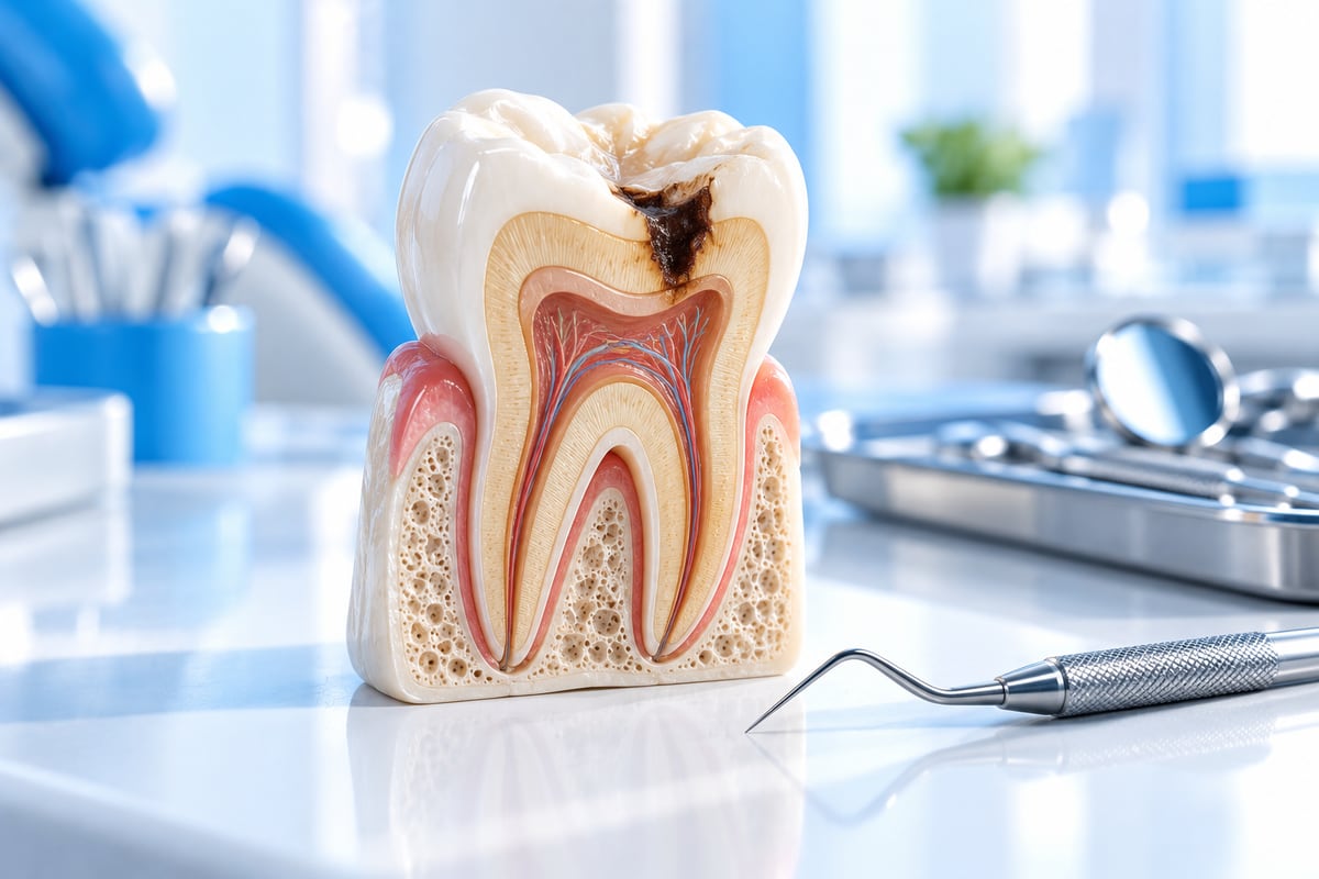
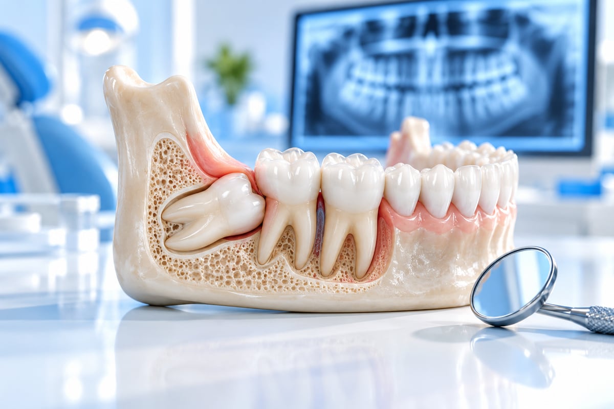
Wisdom teeth are the last to appear, usually between seventeen and twenty-five. The human jaw has become smaller through evolution while the number of teeth stayed the same, so there is often not enough room at the back for four extra molars. The result is familiar to millions: a tooth trying to erupt into a space that cannot hold it, becoming partially or fully impacted, followed by recurring pain and infection.
Types of impaction
- Mesial: the most common — the tooth tilts forward against the second molar, trapping food and causing decay in a healthy tooth.
- Vertical: upright but without room to erupt; often stays quiet for years.
- Horizontal: lying on its side and pressing against the neighbouring root — the most troublesome pattern.
- Distal: tilted backwards, hard to clean and usually surgical.
- Buccal or lingual: angled outward or inward, causing repeated cheek or tongue ulceration.
Symptoms that point to the wisdom tooth
Recurrent pressure at the very back of the jaw, a swollen gum flap behind the last tooth, limited mouth opening, a persistent bad taste, and pain radiating to the ear or temple all point to pericoronitis — inflammation under the gum flap covering a partially erupted tooth. The classic pattern is an episode that settles within a week and returns months later, because the cause has not been removed.
When removal is indicated
- Repeated episodes of pericoronitis.
- Decay in the wisdom tooth that cannot be restored predictably.
- Decay or root resorption in the adjacent second molar.
- A cyst or benign lesion around the crown, often found on imaging before any symptom.
- A deep, uncontrollable periodontal pocket behind the second molar.
- Recurrent trauma to the cheek or tongue.
Equally important is when not to operate: a fully erupted, cleanable, functional wisdom tooth needs nothing, and a fully bony impacted tooth with no symptoms and no pathology can be monitored radiographically in a patient who attends regularly.
What the imaging shows
| Imaging | What it shows | When it is used |
|---|---|---|
| Periapical X-ray | One tooth and the bone around it | A specific localised complaint |
| Panoramic | Both jaws, impaction angle, approximate nerve relationship | First assessment of wisdom teeth |
| CBCT (3D) | Exact relationship to the inferior alveolar nerve and sinus, root anatomy in three dimensions | When the panoramic suggests nerve proximity or complex roots |
Where roots sit very close to the nerve canal, a coronectomy — removing the crown and deliberately leaving the roots — may be proposed. It removes the source of infection while keeping the roots away from the nerve, and requires radiographic follow-up.
The procedure and recovery
Removal is usually done under local anaesthesia, with sedation available for anxious patients, and takes twenty to forty-five minutes per tooth. The gum is opened if needed, a limited amount of bone is removed, and the tooth is often sectioned — a deliberate technique that preserves bone and shortens surgery rather than a sign of difficulty. Swelling peaks on day two or three and settles over five to seven days; limited mouth opening improves with gentle exercises from day three. Full aftercare is covered in our guide on what to do after an extraction.
Common myths
Evidence that wisdom teeth cause lower front crowding is weak — crowding also occurs in people who never had them, so it is not on its own a reason for removal. Wisdom teeth do not affect the eye or brain; pain radiates to the ear and temple because of sensory nerve distribution, and chewing-muscle tension can cause temporal headache. Removing all four at once is an option, not a rule.
Upper wisdom teeth: how they differ
Most discussion focuses on the lower jaw because it causes more trouble, but upper wisdom teeth have their own profile. Upper bone is less dense, so removal is usually quicker with less swelling. In return, the roots can sit close to the floor of the maxillary sinus, which makes imaging essential and means a small oro-antral communication is possible — usually closing on its own or with a simple procedure. After such an extraction, avoid forceful nose-blowing for a week.
Wisdom teeth and orthodontics
If the treatment plan requires moving molars backwards, an impacted wisdom tooth blocking that movement is usually removed first. If it sits far away and does not interfere, the decision can wait until treatment ends. Cleaning the back of the mouth is harder with brackets in place, so pericoronitis is more likely during orthodontic treatment — see our orthodontics page for how cases are planned.
During pregnancy
Pericoronitis is common in pregnancy because hormonal changes make gums more reactive. Local cleaning and irrigation are safe at any stage; paracetamol is the usual painkiller and anti-inflammatories are avoided, particularly in the third trimester. Acute infection with swelling or fever must be treated rather than postponed — infection poses more risk to the pregnancy than treatment does. If extraction is needed, the second trimester is preferred.
What the cost depends on
| Factor | Effect on cost |
|---|---|
| Degree of impaction | An erupted tooth is far simpler than a fully bony one |
| Angle of impaction | Horizontal and distal need more time and technique |
| Proximity to the nerve | May require 3D imaging and a different surgical approach |
| Upper or lower jaw | Lower teeth are usually harder and longer |
| Type of anaesthesia | Local alone, or local with sedation |
| Number removed per visit | Combining teeth in one visit can reduce total cost |
This is why a precise quote over the phone is not possible: examination and imaging come first, then a written plan with the full cost before anything begins.
Managing anxiety
- A step-by-step explanation reduces anxiety more than general reassurance.
- An agreed hand signal to pause at any moment restores a sense of control.
- Anaesthesia is tested before work starts and can be topped up at any point.
- Sedation is available for severe anxiety, leaving you relaxed and aware with limited memory of the procedure.
- Splitting the work across two shorter visits suits many patients.
The silent victim: the second molar
The most damaging thing an impacted wisdom tooth does is not the pain you feel but the harm you do not. When it tilts forward against the second molar, a narrow trap forms between them that neither brush nor floss can reach. Decay starts on the back surface of the second molar — the one surface you cannot see in a mirror — and advances silently for years. By the time it hurts, it has often reached the nerve, turning a simple extraction into root canal treatment and a crown, or the loss of a key chewing tooth that could have been saved. This is why early removal is often advised for a mesial impaction locked against the second molar, even without symptoms.
Wisdom tooth or jaw joint?
| Sign | Wisdom tooth | Jaw joint |
|---|---|---|
| Location | Inside the mouth, right at the back | Directly in front of the ear, temple and cheek muscles |
| On pressure | Tender gum or tooth | Tender joint in front of the ear |
| Sound | None | Clicking or popping on opening |
| Swelling | Possible in gum or cheek | Rare — the problem is muscular or articular |
| Timing | Episodes linked to infection | Worse in the morning or after a stressful day |
| Response | Improves with cleaning and local treatment | Improves with rest, heat and reduced jaw load |
Sometimes both are present: repeated infection around a wisdom tooth makes you chew on one side for months, overloading the muscles on the other. A clinical examination with imaging separates the two quickly — there is no reason to live with undiagnosed pain for months when the answer takes half an hour.
Frequently asked questions
What are the symptoms of an impacted wisdom tooth?
Recurrent pain or pressure at the back of the jaw, a swollen gum flap, limited opening, persistent bad taste and pain radiating to the ear. A fully impacted tooth may have no symptoms at all and be found on a routine X-ray.
Does every wisdom tooth need removing?
No. A well-erupted, cleanable tooth is left alone, and an asymptomatic bony impaction with no pathology can be monitored. Removal is indicated for recurrent infection, decay, damage to the neighbouring tooth or a cyst.
How long is recovery?
Most people return to normal activity in two to three days, with swelling settling over five to seven days; bone healing continues silently for months.
What is the risk of numbness?
Temporary numbness is uncommon and usually resolves over weeks to months; permanent numbness is very rare, and 3D imaging before surgery substantially reduces the risk.
Can it be removed while infected?
Often yes, since removing the source treats the infection — but with spreading swelling or limited opening the infection is usually controlled first.
The bottom line
A wisdom tooth is neither an enemy nor a friend by default. The right decision rests on an examination and imaging that define the impaction, the nerve relationship and the state of the second molar — not on other people's stories. If it causes repeated episodes or threatens the tooth in front, earlier removal is kinder and simpler. If it is quiet, healthy and cleanable, regular monitoring is all you need.
This article is general information for education only and does not replace an examination and diagnosis by a dentist. Every case is different, and the right treatment is decided by your dentist after an examination.
Have a question about your own teeth?
Book an examination at Dental Arts and the dentist will check your teeth and calmly explain the options that suit you.